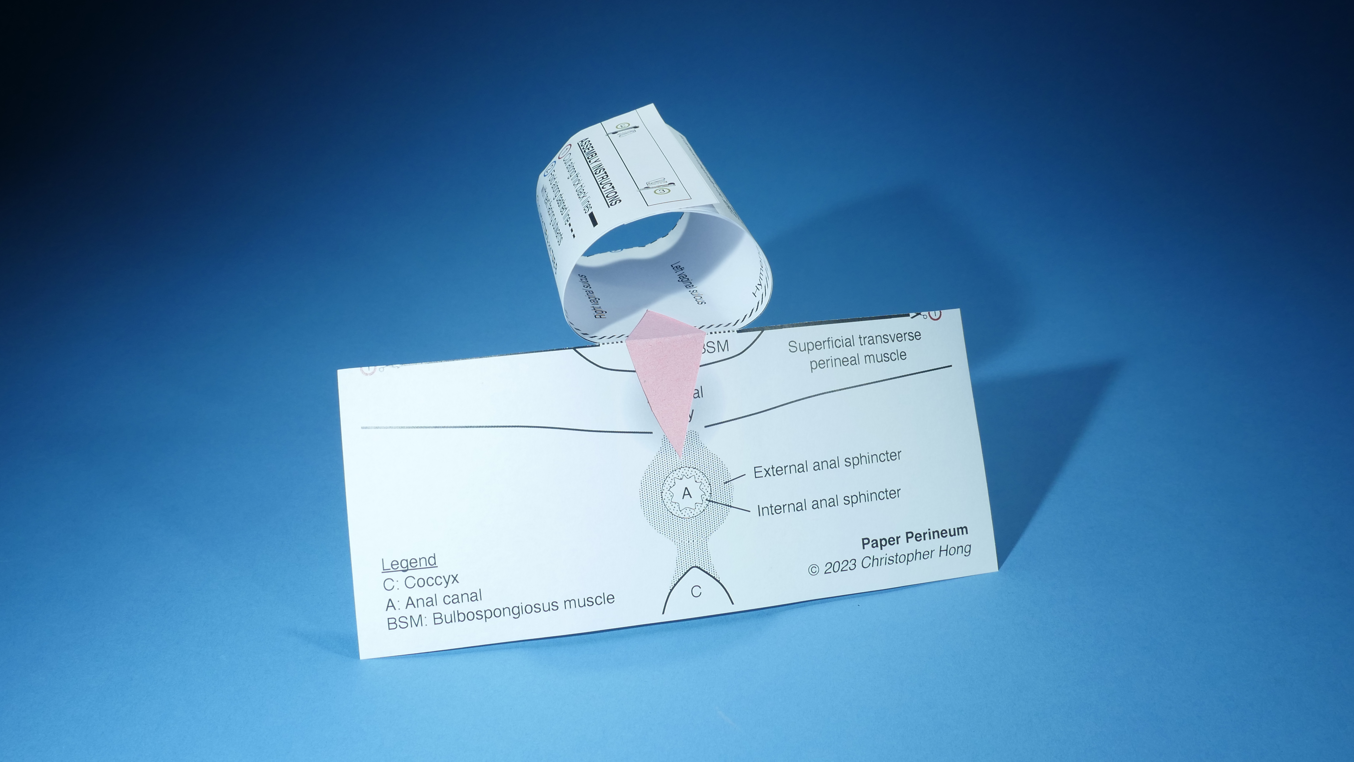
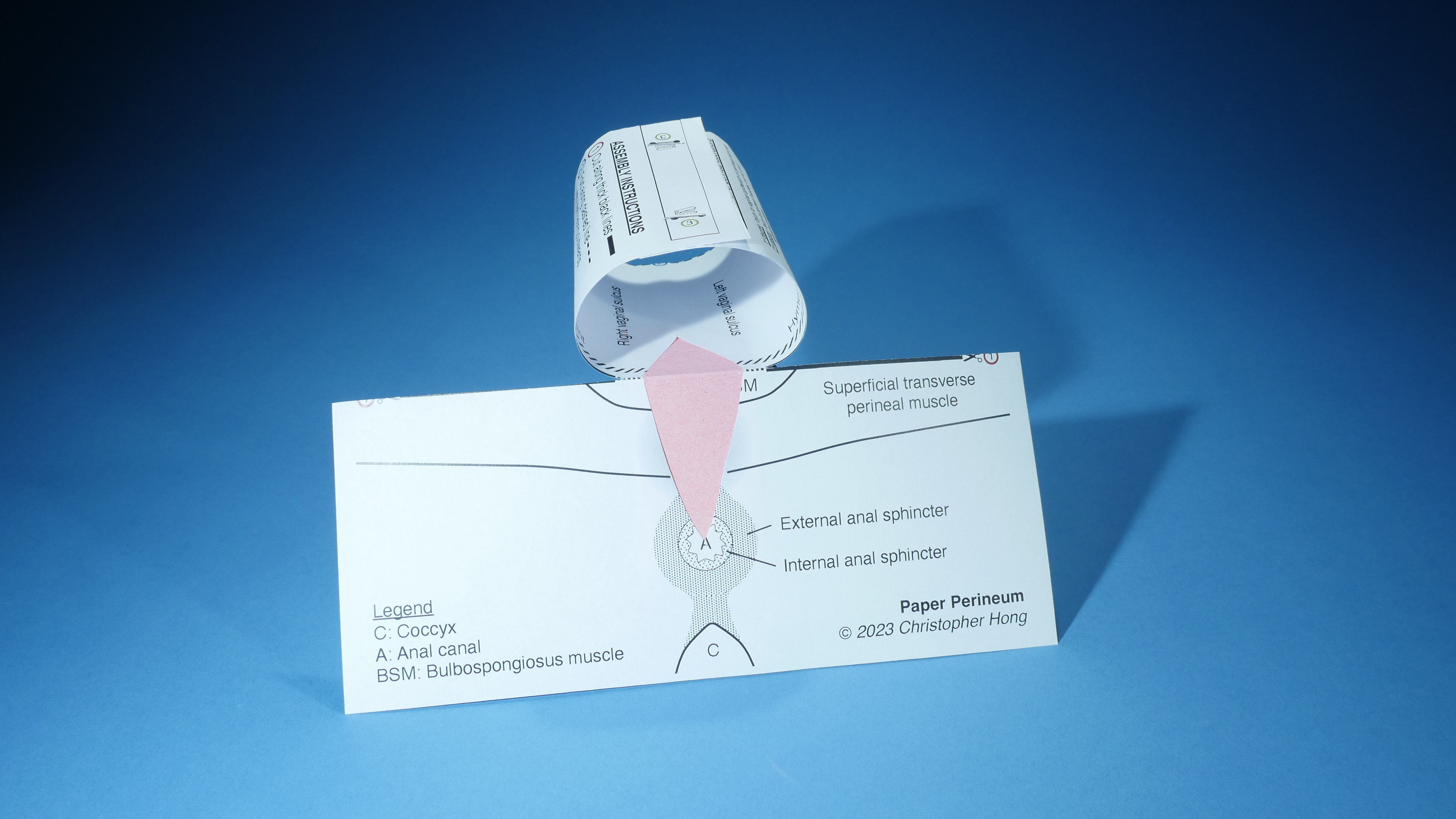
Excise portions of the model to simulate obstetric lacerations and use these models to simulate the three dimensional planes of laceration repairs.
Simulate Lacerations
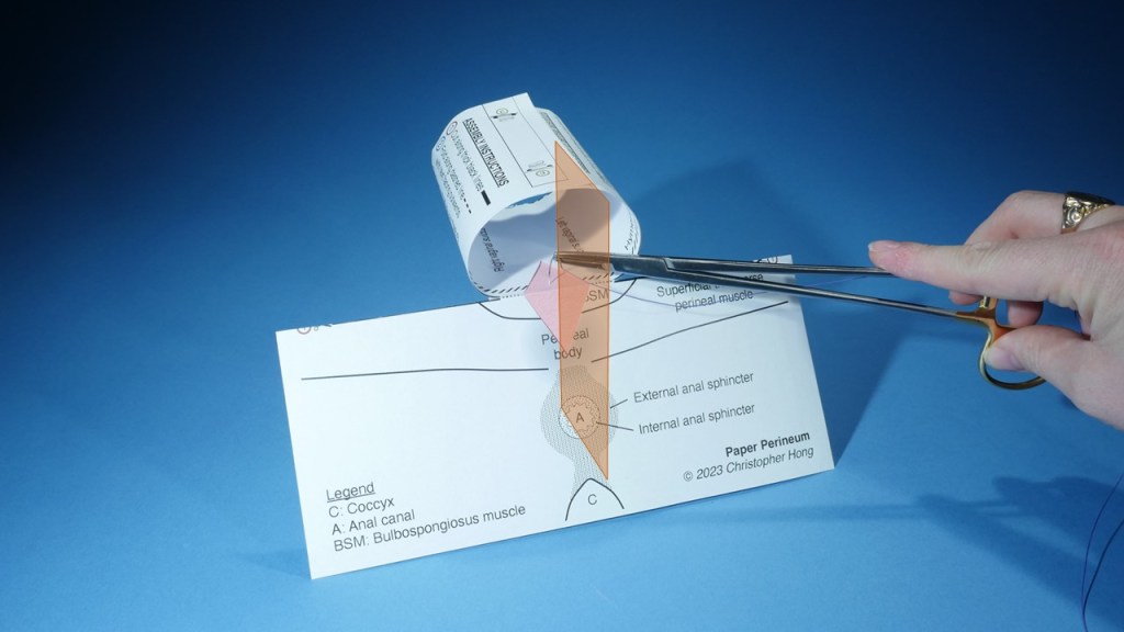
Step 1: Fold the assembled model along the dotted line.
Step 2: Using scissors, excise a triangular portion the model at the junction to the desired depth and shape.
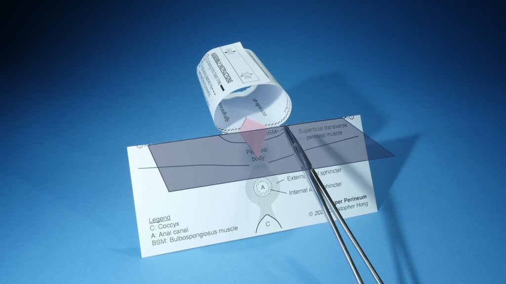
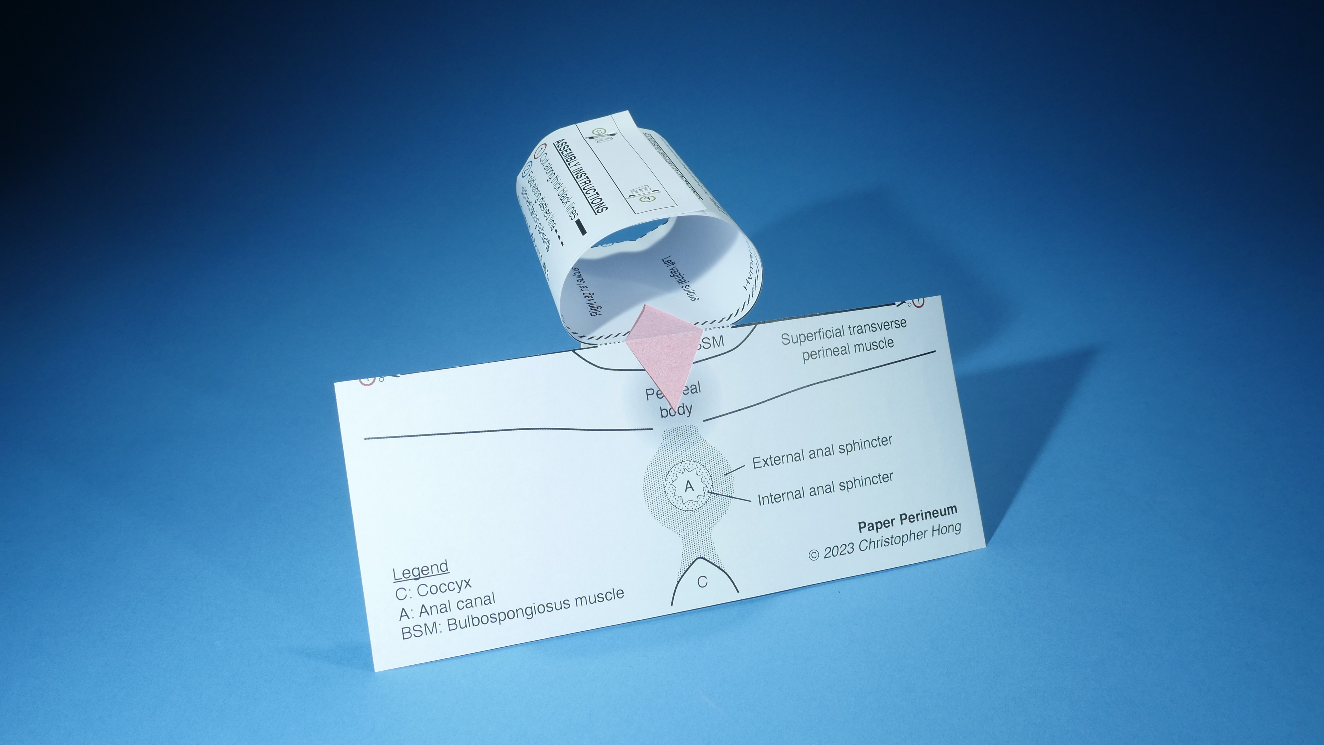
Step 3: Unfold the model.
This creates a diamond shape representing a perineal laceration with vaginal and perineal components. The laceration can be extended deeper into the vagina and towards the anus to simulate additional types of lacerations:
The model can also simulate episiotomies and help learners appreciate how medial episiotomies are more likely to result in anal sphincter injuries:
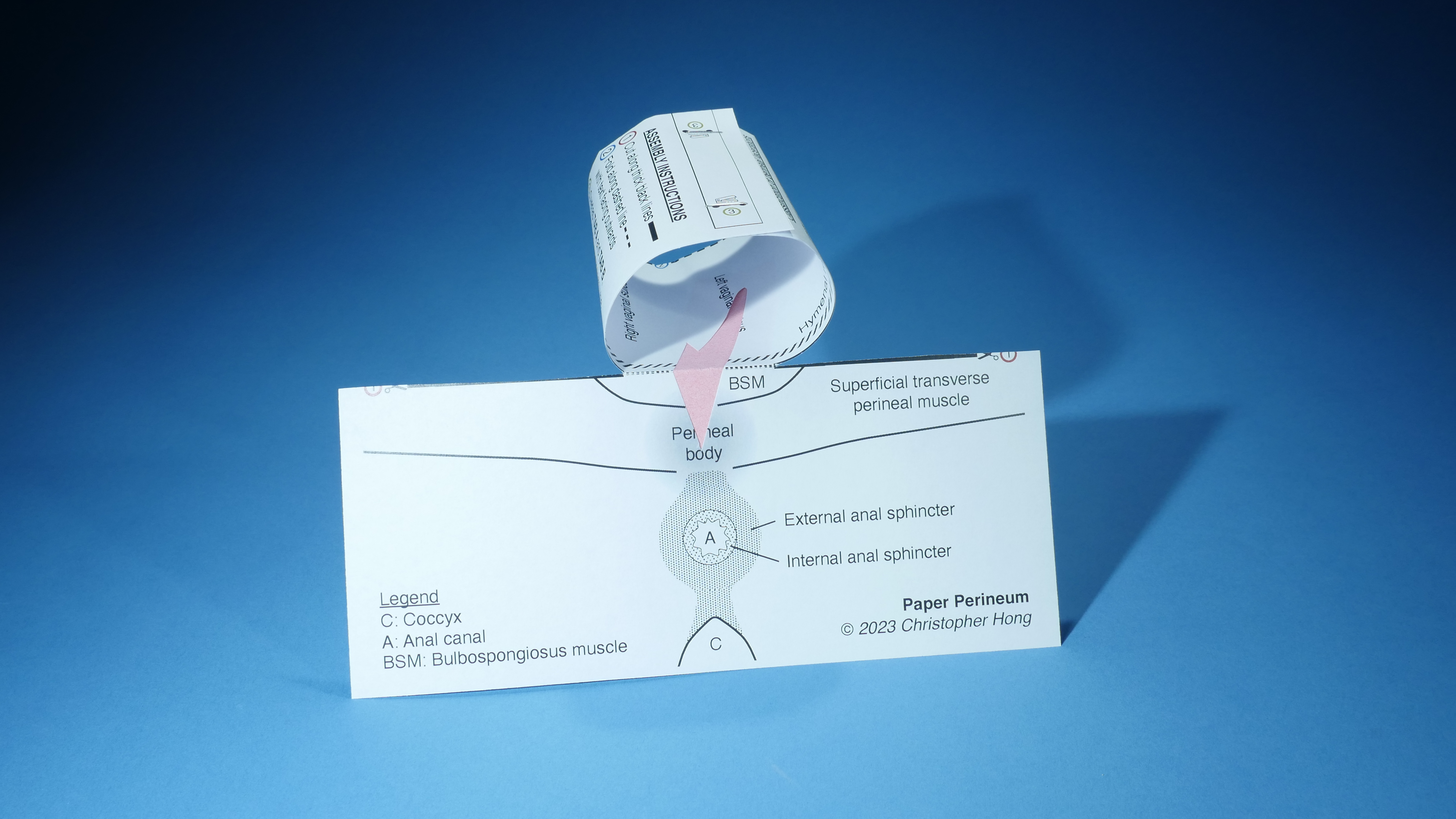
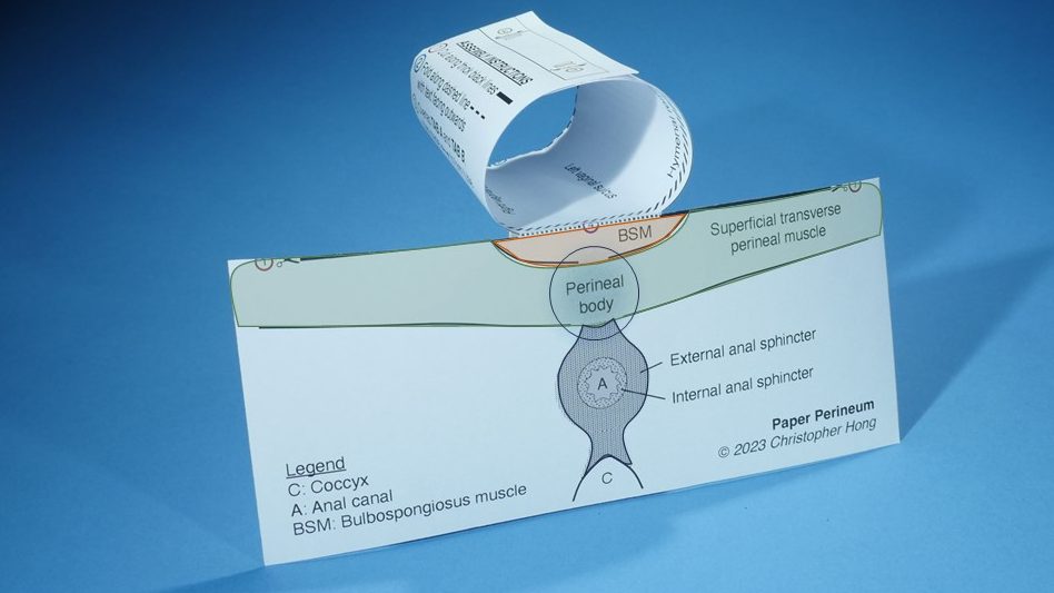
Teach Relevant Anatomy
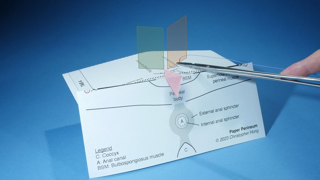
Anatomical Planes
The vaginal canal and perineal surface are oriented perpendicular to each other, and the hymenal remnant acts as a distinguishing landmark between them.
Perineal Structures
The perineal body is formed by intersecting fibers originating from the bulbospongiosus, superficial transverse perineal, and external anal sphincter muscles.
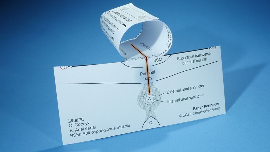
Simulate Laceration Repair
Needle Path
In general, the path of a suture needle should be perpendicular to the edge of the laceration. It may be necessary to adjust the needle’s orientation on the contralateral side of the laceration.